logo
Back to Magazine List

"Is surgery safe for my eyes?" A Must-Read: Guide to 50+ Comprehensive Pre-operative Eye Exams

High-level Eye Exams are a comprehensive diagnostic package designed to enhance the safety and accuracy of all ophthalmic procedures and surgeries, including vision correction, cataract treatment, presbyopia correction, and dry eye management.
Using 3D imaging, AI-assisted analysis, and high-resolution diagnostic devices, the program provides an in-depth evaluation of the entire eye—covering corneal thickness and condition, tear film function, retina, and optic nerve.

PART 1. Vision & Refraction Test
1. Examination Method
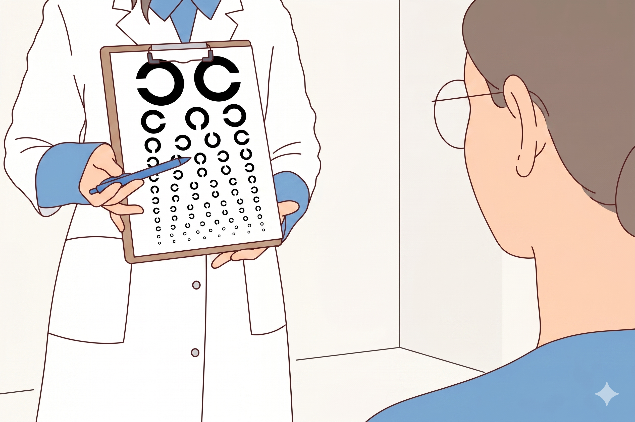
  • Automated refraction testing, distance and near visual acuity tests
  • Measurement of astigmatic axis and refractive power
  • Customized analysis reflecting real-life visual environments
2. Benefits / Effects
  • Accurate assessment of current visual status
  • Identification of the degree of myopia, hyperopia, and astigmatism
  • Determination of the need for correction and surgical eligibility
PART 2. Corneal Topography / Pachymetry

1. Examination Method
  • Analysis of corneal curvature, asymmetry, and astigmatism patterns using high-resolution topography
  • Precise measurement of corneal thickness (CT) via ultrasound or laser-based methods
2. Benefits / Effects
  • Evaluation of eligibility for LASIK, LASEK, or SMILE
  • Early detection of keratoconus or thin cornea
  • Establishment of a safer and more accurate surgical plan

PART 3. Tear Film / Dry Eye Evaluation

1. Examination Method
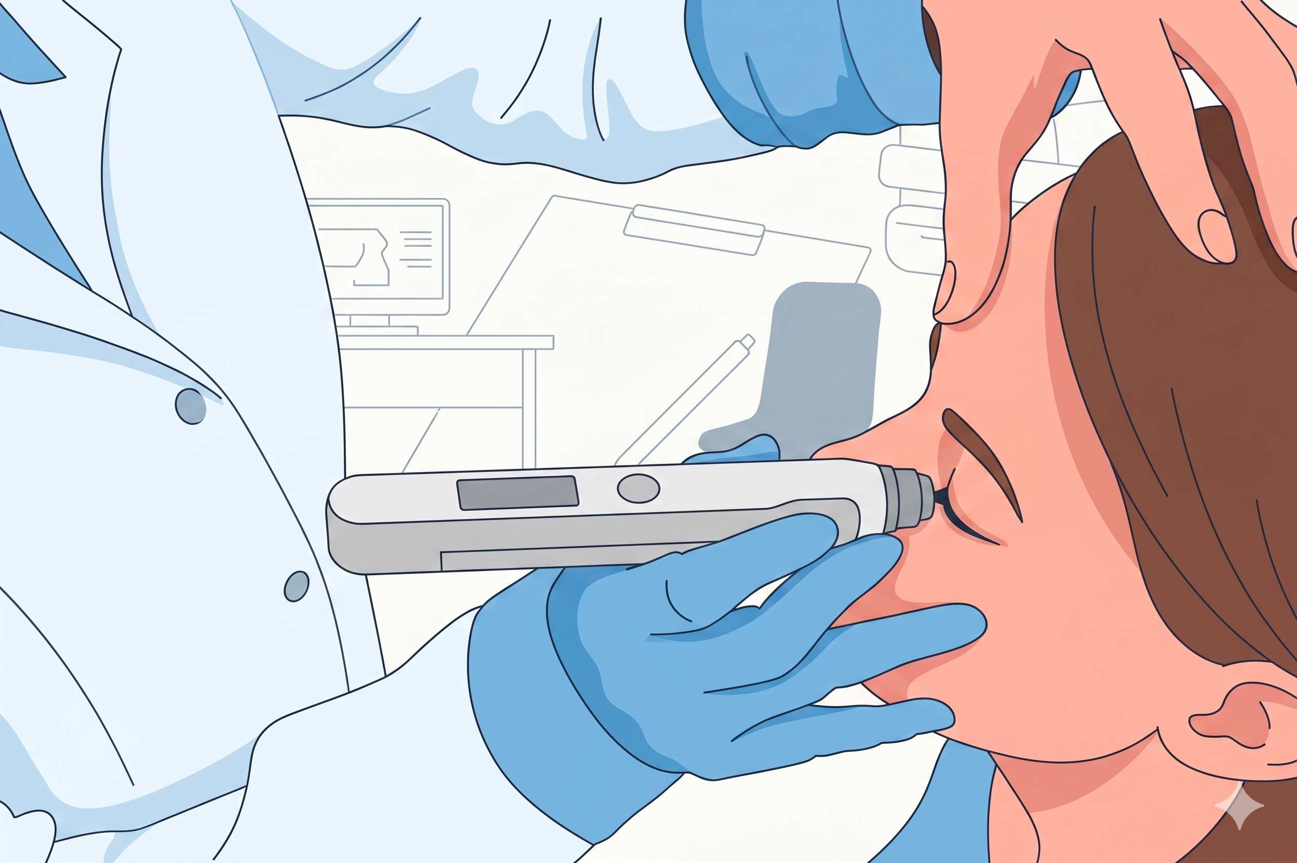
  • Tear film stability test (TBUT), tear secretion assessment, and meibomian gland evaluation
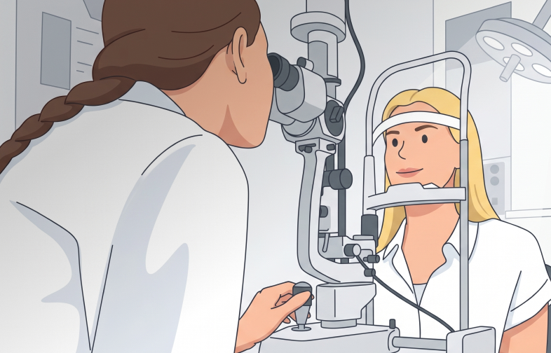
  • Measurement of tear layer quality, thickness, and evaporation patterns using specialized imaging systems
2. Benefits / Effects
  • Accurate identification of the causes of dry eye
  • Determination of the need for preoperative management
  • Personalized treatment direction based on individual tear film characteristics

PART 4. Retina / OCT / Fundus Examination

1. Examination Method
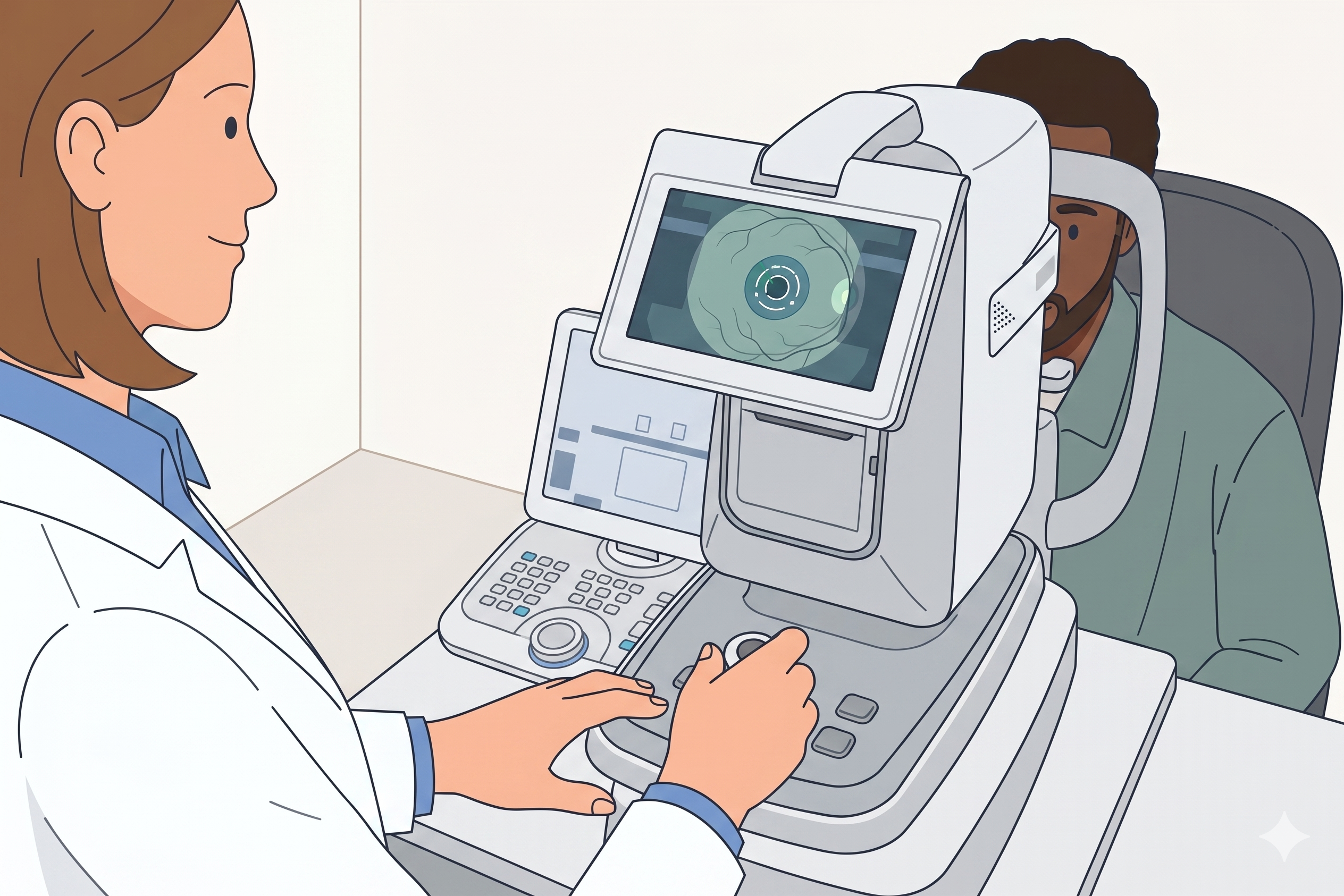
  • Cross-sectional imaging of the retina, optic nerve, and macula using OCT and fundus photography
  • Early detection of vision-threatening diseases such as glaucoma and macular degeneration
2. Benefits / Effects
  • Early identification of risk factors for visual impairment
  • Enhanced safety during procedures and surgeries
  • Essential screening for high-risk patients (family history, high myopia)
Back to Magazine List